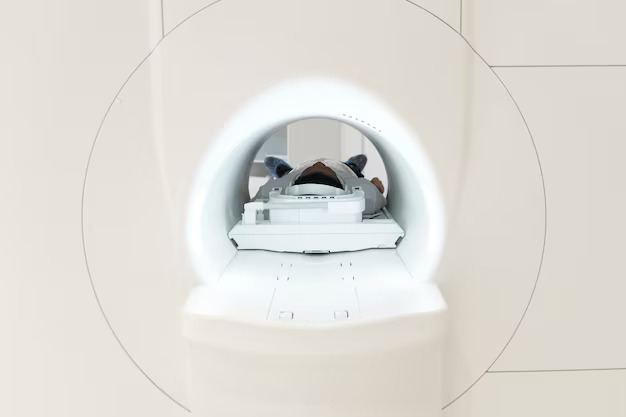
Cone beam CT imaging provides dentists with a three-dimensional view of mouth, jaw, teeth, and nasal cavity. These images contain invaluable clinical information and help reduce the need for invasive procedures, shorten treatment time, and make treatment plans more effective and efficient.
With 3D scans, dentists and dental specialists can now evaluate:
Soft tissue size and location
This is especially important in diagnosing and treating sleep apnea, where soft tissue might be blocking the airways during sleep. These images will also show tumors and irregular growths, and can help your dentist plan for oral surgery.
Location of teeth, including impacted teeth or teeth that haven’t grown in yet
Knowing the exact location and size of your teeth helps in planning treatment for braces or impacted teeth.
Location, size and density of jaw bone
Knowing the location, size, density of your bones will help determine the best plan of action to take if you need an implant or jaw surgery.
When you get a cone beam CT scan, an imaging device rotates around your head. The scanner records between 150 and 600 different X-ray views in under a minute and sends the scans to a computer where a virtual, three-dimensional model is created from the images. The model can be rotated from side to side or up and down, magnified, or viewed from any angle needed.
So not only can your dentist see your entire tooth’s anatomy, they can zoom in to see the condition of your root canal itself. This allows your dentist or dental specialist to prepare your procedure, or examine your health in great detail without you having to sit in an uncomfortable position, or without you even needing to be present.
Like X-rays, CT scans are associated with low amounts of radiation exposure, so it’s important to consider the risk before getting a scan. Most often, the benefits of getting a CT scan outweigh the risks, but it’s particularly important to be cautious for those with preexisting health conditions.


